Breast cancer is the most common type of cancer in American women, according to the American Cancer Society. While women over 50 are most likely to be diagnosed with breast cancer, it can still occur in younger women.
216K
Each year, nearly 216,000 women and 1,500 men learn they have breast cancer.
59K
59,000 women learn they have in situ or noninvasive breast cancer every year.
80%
Over 80% of women diagnosed do not have family history of the disease.
40K
Nearly 40,000 women will die from breast cancer each year.
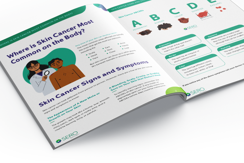
Breast Cancer Symptoms and Risk Factors
Symptoms and signs of breast cancer vary from person to person. If you are experiencing changes to your breasts, it’s important to contact your doctor to determine the cause.
Breast Cancer Symptoms
- Breast pain or discomfort
- Change in breast shape or size
- Nipple discharge
- Skin changes in one or both breasts
- Itchy breasts
- Breast thickening
- Flaking nipple skin
Most women who develop breast cancer do not have known risk factors, but some factors may increase the chance of developing this disease. One of these risk factors is age — the median age of a breast cancer diagnosis is 62 in the United States, according to Susan G. Komen.
Other risk factors include:
- Early onset of menstruation
- Family history of breast cancer
- Hormone replacement therapy with estrogen and progesterone
- Alcohol consumption
- A personal history of breast cancer or prior breast biopsy for benign disease
Diagnosing Breast Cancer
Breast tumors are typically, but not always, painless, so it is important to have any breast or underarm lump checked. Swelling, discoloration, thickening of the skin, or nipple discharge also should be checked immediately.
In some cases, a biopsy to determine if you have breast cancer will be done in an office setting using a needle to remove cells from the lump.
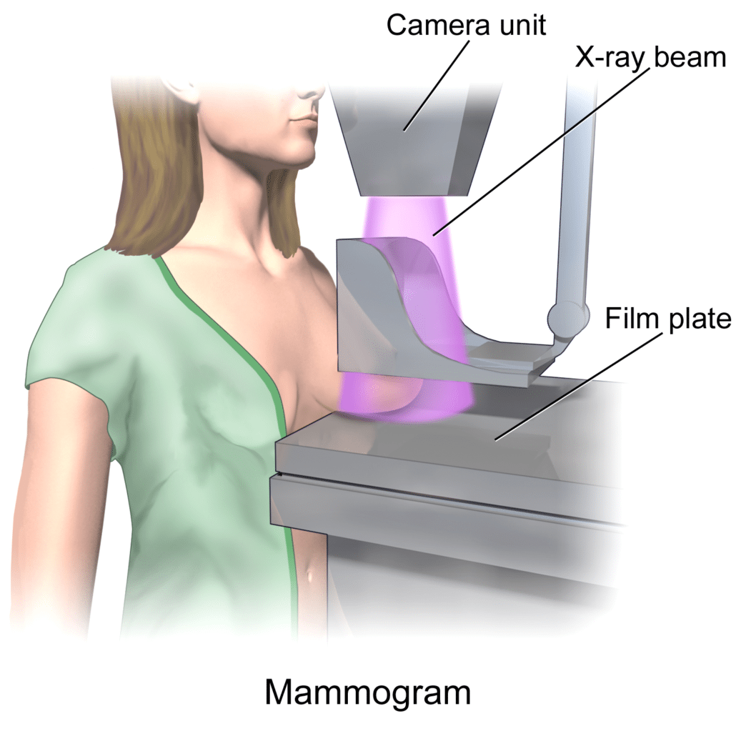
A stereotactic biopsy uses mammography targeting to pinpoint smaller tumors and permit a small amount of tissue to be removed by a needle for diagnosis. Your surgeon may suggest removing the lump to see if you have cancer.
Breast Cancer Treatment
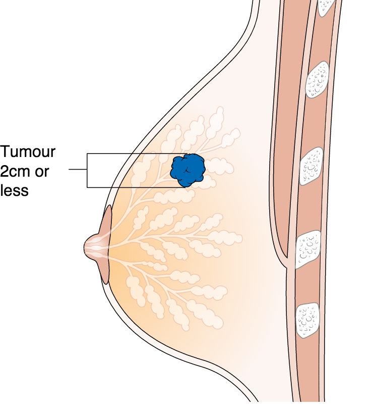
The breast is made up of ducts and lobules surrounded by fatty tissue. Cancer confined within a duct is called ductal carcinoma in situ (DCIS). Lobular carcinoma in situ (LCIS) is cells confined to a lobule. Tumors that break through the wall of the duct or lobule are called infiltrating ductal or infiltrating lobular carcinomas. Inflammatory breast cancer may involve the entire breast with specific skin changes and swelling. Breast cancer treatment often depends on various factors.
If you’re ready to take the next steps in your breast cancer treatment, consider the radiation oncologists at SERO. We have 30+ board-certified physicians in Charlotte, NC and surrounding locations. We are proud and honored to treat patients across the Southeast United States.
Breast Conserving Surgery
Studies have shown that women with early-stage breast cancer who have a lumpectomy to remove the cancer followed by radiation live just as long as women who have a mastectomy and may be preferred by many women. The standard of care after breast-conserving surgery is external beam radiation therapy. Often, this follows chemotherapy.
Your surgeon will perform an operation called a lumpectomy, also called a partial mastectomy, excisional biopsy or tylectomy, to remove the tumor. In some cases, a second operation called a re-excision may be needed if microscopic examination finds tumor cells at or near the edge of the tissue that was removed (called a positive or close margin).
To see if your cancer has spread, your doctor may remove several lymph nodes from under your arm (axilla). If any of these nodes contain cancer cells, more nodes may be removed.
Breast-conserving surgery is not suitable for all breast cancer patients. Talk with your surgeon to see if this is the best procedure for you.
External Beam Radiation
External beam radiation therapy involves a series of daily outpatient treatments to accurately deliver radiation to the breast.
- Painless radiation treatments are delivered in a series of daily sessions. Each treatment will last less than 30 minutes, Monday through Friday, for five to seven weeks.
- The usual course of radiation treats only the breast, although treatment of the lymph nodes around the collarbone or the underarm area is sometimes needed.
- 3-dimensional conformal radiotherapy (3D-CRT) combines multiple radiation treatment fields to deliver very precise doses of radiation to the breast and spare surrounding normal tissue.
- Intensity modulated radiation therapy (IMRT) is a form of 3D-CRT that further modifies the radiation by varying the intensity of the radiation beams. It is currently being studied for treating breast cancer.
- Side effects might include skin irritation, like a mild to moderate sunburn, mild to moderate breast swelling and fatigue.
Partial Breast Irradication
Doctors are studying ways to deliver radiation to only part of the breast. Available in a few clinics for a very select group of patients, these techniques are used after a lumpectomy to deliver radiation to the tumor site rather than the entire breast.
Breast brachytherapy involves placing flexible plastic tubes called catheters or a balloon into the breast. Over one to five days, the catheters or the balloon are connected to a brachytherapy machine so high doses of radiation can treat the nearby breast tissue. MammoSite™ is an example of a balloon catheter device used in this procedure.
Other techniques include 3-D conformal partial breast irradiation and intra-operative radiation therapy (IORT).
The long-term results of these techniques are still being studied. Talk with your radiation oncologist if you would like more information.
After Mastectomy Radiation
In cases where the breast is surgically removed, your doctor may suggest radiation therapy for the chest wall and nearby lymph node areas.
Whether or not radiation therapy should be used after removal of the breast depends on several factors, including the number of lymph nodes involved, tumor size and surgical margins.
Share This Page