Stages of Intraocular (Uveal) Melanoma

Posted on October 16, 2015 in Skin Cancer

Written by Dr. Corso

Learn more about the author

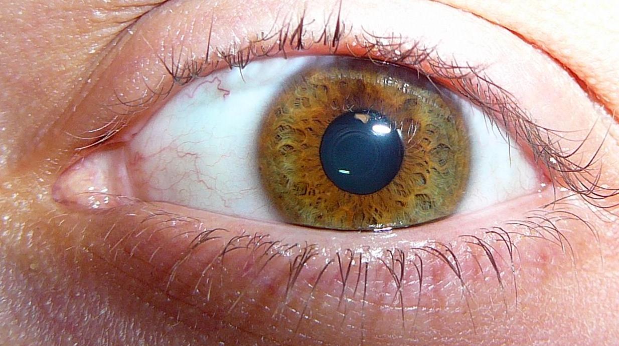
close up of eye with Intraocular (Uveal) Melanoma

After intraocular melanoma has been diagnosed, tests are done to find out if cancer cells have spread to other parts of the body.

The process used to find out if cancer has spread to other parts of the body is calledstaging. The information gathered from the staging process determines the stage of the disease. It is important to know the stage in order to plan treatment.

The following tests and procedures may be used in the staging process:

arrow_forward

Blood chemistry studies

A procedure in which a blood sample is checked to measure the amounts of certain substances released into the blood by organs and tissues in the body. An unusual (higher or lower than normal) amount of a substance can be a sign of disease.

arrow_forward

Liver function tests

 A procedure in which a blood sample is checked to measure the amounts of certain substances released into the blood by the liver. A higher than normal amount of a substance can be a sign the cancer has spread to the liver.

arrow_forward

Ultrasound exam

A procedure in which high-energy sound waves (ultrasound) are bounced off internal tissues or organs, such as the liver, and make echoes. The echoes form a picture of body tissues called a sonogram.

arrow_forward

Chest x-ray

 An x-ray of the organs and bones inside the chest. An x-ray is a type of energy beam that can go through the body and onto film, making a picture of areas inside the body.

arrow_forward

MRI (magnetic resonance imaging)

A procedure that uses a magnet, radio waves, and a computer to make a series of detailed pictures of areas inside the body. This procedure is also called nuclear magnetic resonance imaging (NMRI).

arrow_forward

CT scan (CAT scan)

A procedure that makes a series of detailed pictures of areas inside the body, such as the chest, abdomen, or pelvis, taken from different angles. The pictures are made by a computer linked to an x-ray machine. A dye may be injected into a vein or swallowed to help the organs or tissues show up more clearly. This procedure is also called computed tomography, computerized tomography, or computerized axial tomography.

arrow_forward

PET scan (positron emission tomography scan)

A procedure to find malignanttumor cells in the body. A very small amount of radioactive glucose (sugar) is injected into a vein. The PET scanner rotates around the body and makes a picture of where glucose is being used in the body. Malignant tumor cells show up brighter in the picture because they are more active and take up more glucose than normal cells do. Sometimes a PET scan and a CT scan are done at the same time. If there is any cancer, this increases the chance that it will be found.

The following sizes are used to describe intraocular melanoma:

Small

The tumor is 5 to 16 millimeters in diameter and from 1 to 3 millimeters thick.

Medium

The tumor is 16 millimeters or smaller in diameter and from 3.1 to 8 millimeters thick.

Large

The tumor is:

  • more than 8 millimeters thick and any diameter; or
  • at least 2 millimeters thick and more than 16 millimeters in diameter.

Though most intraocular melanoma tumors are raised, some are flat. These diffuse tumors grow widely across the uvea.

There are three ways that cancer spreads in the body.

Cancer can spread through tissue, the lymph system, and the blood:

  • Tissue. The cancer spreads from where it began by growing into nearby areas.
  • Lymph system. The cancer spreads from where it began by getting into the lymph system. The cancer travels through the lymph vessels to other parts of the body.
  • Blood. The cancer spreads from where it began by getting into the blood. The cancer travels through the blood vessels to other parts of the body.

Cancer may spread from where it began to other parts of the body.

When cancer spreads to another part of the body, it is called metastasis. Cancer cells break away from where they began (the primary tumor) and travel through the lymph system or blood.

  • Lymph system. The cancer gets into the lymph system, travels through the lymph vessels, and forms a tumor (metastatic tumor) in another part of the body.
  • Blood. The cancer gets into the blood, travels through the blood vessels, and forms a tumor (metastatic tumor) in another part of the body.

The metastatic tumor is the same type of cancer as the primary tumor. For example, if intraocular melanoma spreads to the liver, the cancer cells in the liver are actually intraocular melanoma cells. The disease is metastatic intraocular melanoma, not liver cancer.

There are two staging systems for intraocular melanoma.

Intraocular melanoma has two staging systems. The staging system used depends on where in the eye the cancer first formed:

  • Iris.
  • Ciliary body and choroid.

If intraocular melanoma spreads to the optic nerve or nearby tissue of the eye socket, it is called extraocular extension.

The following stages are used for intraocular melanoma of the iris:

Stage I

In stage I, the tumor is in the iris only and is not more than one fourth the size of the iris.

Stage II

Stage II is divided into stages IIA and IIB.

  • In stage IIA, the tumor:
    • is in the iris only and is more than one fourth the size of the iris; or
    • is in the iris only and has caused glaucoma; or
    • has spread next to and/or into the ciliary body, choroid, or both. The tumor has caused glaucoma.
  • In stage IIB, the tumor has spread next to and/or into the ciliary body, choroid, or both, and has also spread into the sclera. The tumor has caused glaucoma.

Stage III

Stage III is divided into stages IIIA and IIIB.

  • In stage IIIA, the tumor has spread through the sclera to the outside of the eyeball. The part of the tumor outside the eyeball is not more than 5 millimeters thick.
  • In stage IIIB, the tumor has spread through the sclera to the outside of the eyeball. The part of the tumor outside the eyeball is more than 5 millimeters thick.

Stage IV

In stage IV, the tumor may be any size and has spread:

  • to nearby lymph nodes; or
  • to other parts of the body, such as the liver, lung, or bone, or to areas under the skin.

The following stages are used for intraocular melanoma of the ciliary body and choroid:

Intraocular melanoma of the ciliary body and choroid is grouped into four size categories. The category depends on how wide and thick the tumor is. Category 1 tumors are the smallest and category 4 tumors are the biggest.

Category 1:

  • The tumor is not more than 12 millimeters wide and not more than 3 millimeters thick; or
  • the tumor is not more than 9 millimeters wide and 3.1 to 6 millimeters thick.

Category 2:

  • The tumor is 12.1 to 18 millimeters wide and not more than 3 millimeters thick; or
  • the tumor is 9.1 to 15 millimeters wide and 3.1 to 6 millimeters thick; or
  • the tumor is not more than 12 millimeters wide and 6.1 to 9 millimeters thick.

Category 3:

  • The tumor is 15.1 to 18 millimeters wide and 3.1 to 6 millimeters thick; or
  • the tumor is 12.1 to 18 millimeters wide and 6.1 to 9 millimeters thick; or
  • the tumor is 3.1 to 18 millimeters wide and 9.1 to 12 millimeters thick; or
  • the tumor is 9.1 to 15 millimeters wide and 12.1 to 15 millimeters thick.

Category 4:

  • The tumor is more than 18 millimeters wide and may be any thickness; or
  • the tumor is 15.1 to 18 millimeters wide and more than 12 millimeters thick; or
  • the tumor is 12.1 to 15 millimeters wide and more than 15 millimeters thick.

Stage I

In stage I, the tumor is size category 1 and is in the choroid only.

Stage II

Stage II is divided into stages IIA and IIB.

  • In stage IIA, the tumor:
    • is size category 1 and has spread to the ciliary body; or
    • is size category 1 and has spread through the sclera to the outside of the eyeball. The part of the tumor outside the eyeball is not more than 5 millimetersthick. The tumor may have spread to the ciliary body; or
    • is size category 2 and is in the choroid only.
  • In stage IIB, the tumor:
    • is size category 2 and has spread to the ciliary body; or
    • is size category 3 and is in the choroid only.

Stage III

Stage III is divided into stages IIIA, IIIB, and IIIC.

  • In stage IIIA, the tumor:
    • is size category 2 and has spread through the sclera to the outside of the eyeball. The part of the tumor outside the eyeball is not more than 5 millimetersthick. The tumor may have spread to the ciliary body; or
    • is size category 3 and has spread to the ciliary body; or
    • is size category 3 and has spread through the sclera to the outside of the eyeball. The part of the tumor outside the eyeball is not more than 5 millimeters thick. The tumor has not spread to the ciliary body; or
    • is size category 4 and is in the choroid only.
  • In stage IIIB, the tumor:
    • is size category 3 and has spread through the sclera to the outside of the eyeball. The part of the tumor outside the eyeball is not more than 5 millimeters thick. The tumor has spread to the ciliary body; or
    • is size category 4 and has spread to the ciliary body; or
    • is size category 4 and has spread through the sclera to the outside of the eyeball. The part of the tumor outside the eyeball is not more than 5 millimeters thick. The tumor has not spread to the ciliary body.
  • In stage IIIC, the tumor:
    • is size category 4 and has spread through the sclera to the outside of the eyeball. The part of the tumor outside the eyeball is not more than 5 millimetersthick. The tumor has spread to the ciliary body; or
    • may be any size and has spread through the sclera to the outside of the eyeball. The part of the tumor outside the eyeball is more than 5 millimeters thick. The tumor has not spread to the ciliary body.

Stage IV

In stage IV, the tumor may be any size and has spread:

  • to nearby lymph nodes; or
  • to other parts of the body, such as the liver, lung, or bone, or to areas under the skin.