Intraocular (Eye) Melanoma

Posted on October 12, 2015 in Cancer

Written by Dr. Moeller

Learn more about the author

What is Intraocular (Eye) Melanoma?

Anatomy of the Eye graphic

Intraocular melanoma is a rare disease in which cancer forms in melanocytes in the eye. Melanocytes are cells that make melanin (the pigment that gives skin and eyes their color).

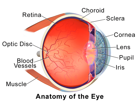
Intraocular melanoma begins in the uvea. The uvea has three parts. The iris is the colored area at the front of the eye. The ciliary body is a ring of muscle tissue that changes the size of the pupil and the shape of the lens. The choroid is a layer of blood vessels that brings oxygen and nutrients to the eye. Most intraocular melanomas begin in the choroid.

There may be no early signs or symptoms of intraocular melanoma. It is sometimes found during an eye exam.

Risk factors for intraocular melanoma include having fair skin that freckles and burns easily, does not tan, or tans poorly and having blue, green, or other light-colored eyes.

  • Being older and having fair skin may increase the risk of intraocular melanoma.
  • Signs of intraocular melanoma include blurred vision or a dark spot on the iris.
  • Tests that examine the eye are used to help detect (find) and diagnose intraocular melanoma.
  • A biopsy of the tumor is rarely needed to diagnose intraocular melanoma.
  • Certain factors affect prognosis (chance of recovery) and treatment options.

Intraocular melanoma is a disease in which malignant (cancer) cells form in the tissues of the eye.

Intraocular melanoma begins in the middle of three layers of the wall of the eye. The outer layer includes the white sclera (the “white of the eye”) and the clear cornea at the front of the eye. The inner layer has a lining of nerve tissue, called the retina, which senses light and sends images along the optic nerve to the brain.

The middle layer, where intraocular melanoma forms, is called the uvea or uveal tract, and has three main parts:

  • Iris

    The iris is the colored area at the front of the eye (the “eye color”). It can be seen through the clear cornea. The pupil is in the center of the iris and it changes size to let more or less light into the eye. Intraocular melanoma of the iris is usually a smalltumor that grows slowly and rarely spreads to other parts of the body.

  • Ciliary body

    The ciliary body is a ring of tissue with muscle fibers that change the size of the pupil and the shape of the lens. It is found behind the iris. Changes in the shape of the lens help the eye focus. The ciliary body also makes the clear fluid that fills the space between the cornea and the iris. Intraocular melanoma of the ciliary body is often larger and more likely to spread to other parts of the body than intraocular melanoma of the iris.

  • Choroid

    The choroid is a layer of blood vessels that bring oxygen and nutrients to the eye. Most intraocular melanomas begin in the choroid. Intraocular melanoma of the choroid is often larger and more likely to spread to other parts of the body than intraocular melanoma of the iris.

Signs

Signs of intraocular melanoma include blurred vision or a dark spot on the iris.

Intraocular melanoma may not cause early signs or symptoms. It is sometimes found during a regular eye exam when the doctor dilates the pupil and looks into the eye. Signs and symptoms may be caused by intraocular melanoma or by other conditions. Check with your doctor if you have any of the following:

  • Blurred vision or other change in vision.
  • Floaters (spots that drift in your field of vision) or flashes of light.
  • A dark spot on the iris.
  • A change in the size or shape of the pupil.
  • A change in the position of the eyeball in the eye socket.

Diagnosis

Tests that examine the eye are used to help detect (find) and diagnose intraocular melanoma.

The following tests and procedures may be used:

  • Physical exam and history : An exam of the body to check general signs of health, including checking for signs of disease, such as lumps or anything else that seems unusual. A history of the patient’s health habits and past illnesses and treatments will also be taken.
  • Eye exam with dilated pupil: An exam of the eye in which the pupil is dilated (enlarged) with medicated eye drops to allow the doctor to look through the lens and pupil to the retina. The inside of the eye, including the retina and the optic nerve, is checked. Pictures may be taken over time to keep track of changes in the size of the tumor. There are several types of eye exams:
    • Ophthalmoscopy : An exam of the inside of the back of the eye to check the retina and optic nerve using a small magnifying lens and a light.
    • Slit-lamp biomicroscopy : An exam of the inside of the eye to check the retina, optic nerve, and other parts of the eye using a strong beam of light and amicroscope.
    • Gonioscopy : An exam of the front part of the eye between the cornea and iris. A special instrument is used to see if the area where fluid drains out of the eye is blocked.
  • Ultrasound exam of the eye: A procedure in which high-energy sound waves (ultrasound) are bounced off the internal tissues of the eye to make echoes. Eye drops are used to numb the eye and a small probe that sends and receives sound waves is placed gently on the surface of the eye. The echoes make a picture of the inside of the eye and the distance from the cornea to the retina is measured. The picture, called asonogram, shows on the screen of the ultrasound monitor.
  • High-resolution ultrasound biomicroscopy : A procedure in which high-energy sound waves (ultrasound) are bounced off the internal tissues of the eye to make echoes. Eye drops are used to numb the eye and a small probe that sends and receives sound waves is placed gently on the surface of the eye. The echoes make a more detailed picture of the inside of the eye than a regular ultrasound. The tumor is checked for its size, shape, and thickness, and for signs that the tumor has spread to nearby tissue.
  • Transillumination of the globe and iris: An exam of the iris, cornea, lens, and ciliary body with a light placed on either the upper or lower lid.
  • Fluorescein angiography : A procedure to look at blood vessels and the flow of bloodinside the eye. An orange fluorescent dye (fluorescein) is injected into a blood vessel in the arm and goes into the bloodstream. As the dye travels through blood vessels of the eye, a special camera takes pictures of the retina and choroid to find any areas that are blocked or leaking.
  • Indocyanine green angiography: A procedure to look at blood vessels in the choroid layer of the eye. A green dye (indocyanine green) is injected into a blood vessel in the arm and goes into the bloodstream. As the dye travels through blood vessels of the eye, a special camera takes pictures of the retina and choroid to find any areas that are blocked or leaking.
  • Ocular coherence tomography : An imaging test that uses light waves to take cross-section pictures of the retina, and sometimes the choroid, to see if there is swelling or fluid beneath the retina.

A biopsy of the tumor is rarely needed to diagnose intraocular melanoma.

A biopsy is the removal of cells or tissues so they can be viewed under a microscope to check for signs of cancer. Rarely, a biopsy of the tumor is needed to diagnose intraocular melanoma. Tissue that is removed during a biopsy or surgery to remove the tumor may be tested to get more information about prognosis and which treatment options are best.

The following tests may be done on the sample of tissue:

  • Cytogenetic analysis: A laboratory test in which cells in a sample of tissue are viewed under a microscope to look for certain changes in the chromosomes.
  • Gene expression profiling : A laboratory test in which cells in a sample of tissue are checked for certain types of RNA.

A biopsy may result in retinal detachment (the retina separates from other tissues in the eye). This can be repaired by surgery.

Certain factors affect prognosis (chance of recovery) and treatment options.

The prognosis (chance of recovery) and treatment options depend on the following:

  • How the melanoma cells look under a microscope.
  • The size and thickness of the tumor.
  • The part of the eye the tumor is in (the iris, ciliary body, or choroid).
  • Whether the tumor has spread within the eye or to other places in the body.
  • Whether there are certain changes in the genes linked to intraocular melanoma.
  • The patient’s age and general health.
  • Whether the tumor has recurred (come back) after treatment.